
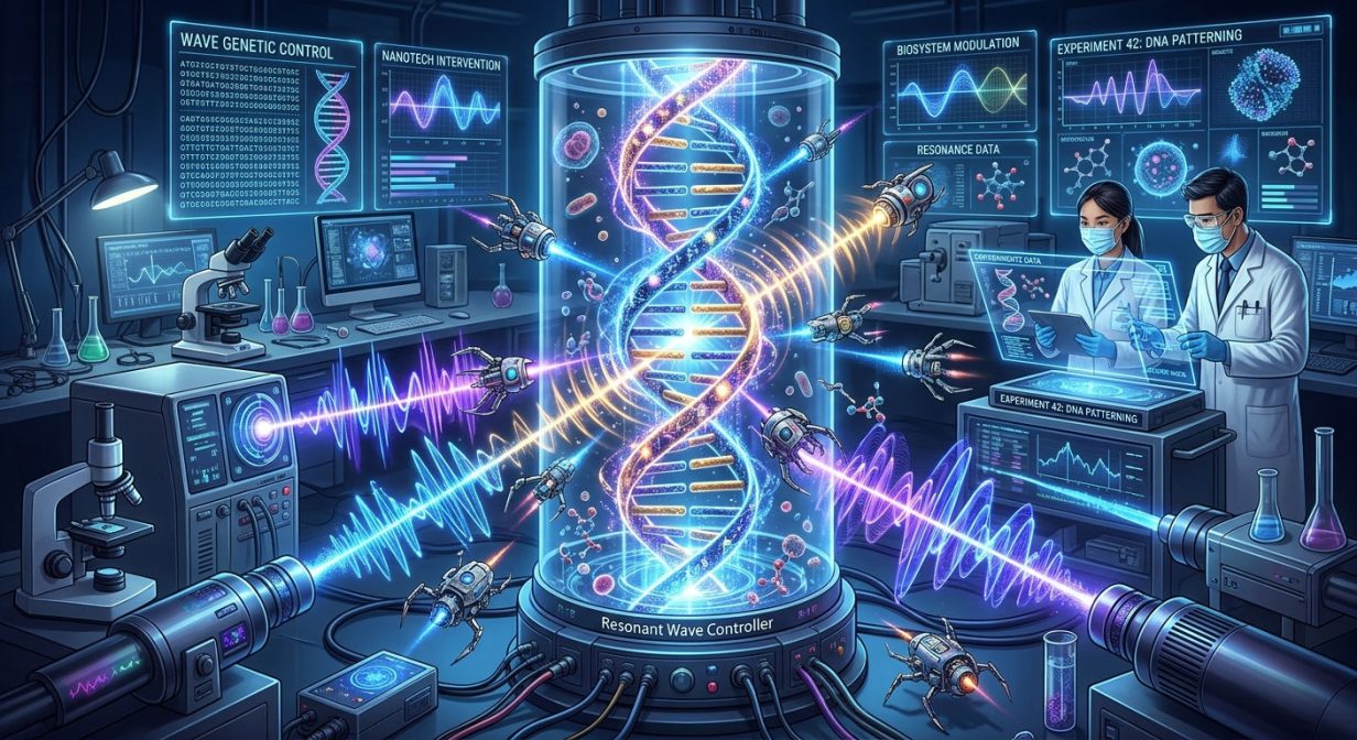
Wave Genetic Nanotechnologies for Biosystem Control: Theory and Experiments
G.G. Tertyshny, P.P. Garyaev
New Medical Technologies, No. 7, pp. 49–64 (2007)
English version, prepared for online publication
Abstract
In the present work we develop our previously proposed theory [3, 4, 7–13, 25, 26, 29–31, and others] of external quantum control and self-control of organisms in vitro – in vivo by means of bioholographic information. The main publications in this direction are collected on the website “Wave Genetics” [35]. The current stage of the theory concerns bio-sign polarization of laser light — that is, holography — which occurs when matrix genetic nanostructures (donors) are scanned (read out) by a beam from a Helium-Neon (HeNe) laser, 632.8 nm (red), operating with the MBER effect. Biosystems are also capable of scanning and correcting (computing) themselves by means of their own coherent chromosome continuum emissions in the 250–800 nm range. Our artificial approach merely reproduces endogenous nanotechnologies in vitro. In such computing — whether in a living organism or in its artificial human-made repetition — a pool of broad-spectrum wave information is generated, which organisms use for their own regulation, and which we harness for purposeful positive control of the metabolism of biosystems.
A mathematical model is presented of polarization-dynamic acts of selected metabolic change in biosystems by means of laser holography-computing in vitro – in vivo. General mechanisms of such acts of natural and artificial control of biosystems are discussed, together with details of the method and apparatus used in practical work in this direction. From the standpoint of theory and its application, we review several of our earlier experimental studies of this kind, which we believe support our models of wave genetic functions of biosystems.
Introductory Remarks
The concept and term “holography” derive from two Greek words meaning “whole” and “image.” Until recently, the essence of holography amounted to a technical method for producing complete spatial (three-dimensional) and spatio-temporal (four-dimensional) images of an object. Now the concept of holography has been radically expanded and extended to the structures and functions of the cerebral cortex [32] and of the genetic apparatus of organisms. When we speak of genetic memory, this means that the chromosome continuum, acting as a quantum biocomputer, operates with four-dimensional wave images of its own dynamic structure for strategic control of metabolism [3, 4, 7–13, 25, 26, 29–31].
With a phase (transparent) structure of the holographed object, a complete and detailed image of it is obtained throughout the surrounding space. The holographic method was first proposed by D. Gabor in 1948 and substantially advanced by Soviet scientists. The method is based on the interference of coherent radiation of any nature. For example, a “signal” wave scattered by the object is directed simultaneously with a “reference” (etalon) wave from the same light source, bypassing the object, onto a photographic plate. The interference pattern arising from these waves — containing complete information about the object — is recorded on the photosensitive surface; it is called a hologram. When the hologram (or a fragment of it) is illuminated by the reference wave, a volumetric image of the entire object becomes visible. Holography is widely used in physics and in various branches of technology (in particular, for pattern recognition and information encoding), in acoustics (for detecting internal defects in critical metal structures, for example at nuclear power stations), and so on. Holography holds great promise for volumetric cinema and television [1].
The present work (like a number of others [2–13, 21, 36–40]) continues investigations begun under the guidance of I.V. Prangishvili, who warmly supported the then-new hypothesis (1997–2001) of holographic properties of biosystems and of the possibility of holographic control over them. By holographic control we mean the alteration of metabolism and of cell structure as a result of governing acoustic, optical, or electromagnetic image-bearing influences.
In the control of biological systems, holographic information is transferred from a donor to a recipient. In the course of laser-holographic experiments on plants in 1997, the phenomenon of holographic translation of information from donor to recipient was physically and mathematically substantiated [9–13, 21]. The essence of this phenomenon is the passage of a special laser radiation through semi-transparent biological tissues and cells — donors of the wave equivalent of genetic-metabolic information. The donors act here as holographic modulators of the probing light. This modulation is, in fact, polarization-phase holography of the structure and mobile metabolic (including genetic) status of the donor. As a result, a complex dynamic register of four-dimensional image-commands arises, with which the quantum biocomputer we have created operates in order to control recipient organisms. Such an artificial quantum biocomputer essentially reproduces in vitro, in a greatly simplified form, what our genetic apparatus does as a natural DNA-wave biocomputer in vivo [26].
For stable and undistorted in vivo recording of read-out information in a beam flux, we proposed at that time an optical interpretation of cell nuclei as vibration-stable polarization sensor-transformers of a dynamic hologram. The physical basis of such a transformer is the principle of redundant encoding of each amplitude-phase scattering point of the object in the form of polarization quasi-rings of Newton.
In our experiments on pancreatic regeneration in rats [24, 29], vibration-stable transfer of polarization-dynamic holographic information from donor to recipient was carried out. Under sufficiently prolonged and purposeful near-resonant exposure of the recipient, the phenomenon of holographic control of the recipient’s state occurred by means of artificially translated holographic information emanating from the cells and tissues of the donor. As a result, the stem cells of the recipient received an informational impulse to begin differentiation in the direction of post-embryonic morphogenesis, with complete restoration of the pancreas in the rats. We do not know which specific type (or types) of stem cells is engaged here; this is a subject for future research.
In the course of this work it became clear that the main pool of bioholographic information resides in the polarization-dynamic modulations of the Euler angles. This can be explained by the fact that after partial reflection of the laser beam and its passage through each point of the bio-preparation donors, light cones of scattered radiation arise, in which the orthogonally circular polarization emitted by the laser is transformed into its spatially conical distribution. The key event here is the interaction of the scattered radiation of the light cones with the polarization reference wave. The latter is synthesized by the sensor-transformer, which may be represented by associates of polarization-active cell nuclei. In this interaction, spatially distributed polarization quasi-rings of Newton arise.
Living cells are always a metabolically and polarizationally non-stationary medium. Nevertheless, light scattered by such a medium produces quasi-rings of Newton that are practically immobile relative to one another and relative to the origin of coordinates chosen in the space where the donor object is located. This happens because of the relative connectedness (coupling) of the donor’s points with one another. The variable Euler angles are caused by microscopic amplitude oscillations of the donor’s points, corresponding to the dynamic state of the cells of the living biological object. These variable angles are the angles between straight lines tangent to the mobile polarization quasi-rings and the coordinate axes in which the donor’s points are considered.
Moreover, it proved possible to transfer information from the donor into the far zone, where the recipient was located. By far zone, as usual, is meant a distance significantly exceeding the wavelength of the laser probing signal. To understand and realize this process, we developed the concept of cell nuclei as polarization quasi-lenses. The physics and operating principle of such lenses is that they — acting both as polaroids and simultaneously as sources of coherent light (250–800 nm) located in the cytoplasmic cell continuum medium — scan their own and cytoplasmic modulations of polarization. This is a key contribution to the synthesis of bioholograms, and at the same time it is the least explained part of the phenomenology.
The same factors solve the problem of dynamic stability of polarization holograms, which proved especially important for work with living organisms. Under any micro-movements of the laser beam relative to the scanned donor preparation, or of the donor relative to the beam — for example, due to seismic motion of the foundation on which the laser stands and/or due to the non-stationarity of the donor — one and the same relatively stable system of Newton’s polarization rings arises along the donor’s cells. In other words, the polarization bioholographic images formed during laser probing of donors are stable, not blurred, and are therefore recognized by the recipient biosystem as regulatory ones.
In holographic encoding and translation of information we succeeded in solving the problem of additional redundancy preservation. This redundancy is understood here in connection with direct and inverse Fourier transformation, which consists, first, in the formation and registration — from each point of the donor — of quasi-rings of Newton and, second, in their inverse Fourier transformation. The direct Fourier transform yields a system of quasi-rings of Newton for each point of the donor’s cells, while the inverse transform converts these rings into analogous points located in the far zone on the recipient. In the end, redundancy is ensured by the fact that, upon passage through the cell nuclei quasi-lenses, each cellular structure of the donor is transformed into a set of volumetric polarization cones of a standing light wave of intensity. In the case of partial erasure or vibrational blurring of the quasi-rings of Newton corresponding to some point of the recipient, the remaining part of the rings proves necessary and sufficient for correct formation of the corresponding point of the donor.
These are the main distinctions and advantages of the method and apparatus for holographic control of the state of cells of biological systems described in the present work. Thanks to the above solutions, we obtained polarization-dynamic holographic translation of information without its geometric and scale distortions.
We note that for obtaining a hologram, non-coherent radiation may also be used. However, in our case coherent light was used to provide a multitude of feedbacks which, in the final analysis, impart biological activity to the holographically modulated information transmitted over optical, electromagnetic, and acoustic channels. Furthermore, the useful signal emanating from the donor carries a polarization hologram modulated by vibrating quasi-rings of Newton. The modulation of the light flux by the donor tissue is picked up by a quadratic photodetector built into the laser tube. As a result, the modulation is transformed into an alternating electromagnetic signal. It is essential that the modulating vibration of the Newton rings (rings of intensity) reflects the code polarization-phase dynamics of each micro-fragment of the donor — for example, of the liquid-crystal (LC) chromosomes. In turn, the micro-dynamic vibration of these rings (and of straight lines tangent to them) transmits the dynamics of the Euler angles. All of this sign-symbolic dynamics — holographic and “key-lock” — resonantly acts on the recipient biosystem, for example on the LC chromosomes, reprogramming them isomorphically to the donor.
Thus, the polarization-dynamic modulation of the light flux, represented by the quasi-rings of Newton, is transformed, through their motion, into an electromagnetic signal which modulates the carrier frequency of the pulse generator harmonics regulating micro-displacements of the laser resonator mirrors. The maximum depth of modulation of the useful signal falls in the frequency range from 0.5 MHz to 1.5 MHz, which is easily detected and received by practically any medium-wave radio receiver.
In addition, we should note that upon repeated playback of such audio signals, we have detected their biological activity. This applies to many recordings on any medium from both living and non-living donor objects. The results of our observations will be presented in subsequent publications.
Theoretical Justification of the Possibility of Storing, Recording, and Reading Dynamic Polarization Holograms Using Informational Biopolymers
Earlier we carried out successful distant (tens of meters) laser-radiowave transmission of morphogenetic signals from a biodonor (preparations of rat pancreas and spleen) to a biorecipient (rats with type-1 diabetes), which induced regeneration of the pancreas in the bodies of the diseased animals and their complete recovery (control rats died) [24]. This fact requires theoretical-biological and physical explanation, since proof of the possibility of existence of active genetic information in the form of an electromagnetic field has fundamental (worldview) significance.
It is known that the main informational polymers of cells — DNA, RNA, proteins, and many other metabolites — contain asymmetric nitrogen atoms in their composition, in consequence of which these metabolites possess optical activity and polarize light. At the same time it is known that abiogenic nitrogen-containing polymers are capable of recording dynamic polarization holograms with high diffraction efficiency [14]. In this connection it is of interest to consider the informational biopolymers — DNA, RNA, and proteins — as possible holders and substrates for recording polarization-bioholographic information, given that DNA, RNA, and proteins are also nitrogen-containing polymers. It is possible that, by virtue of such similarity, DNA, RNA, and proteins are capable in a special way of absorbing light quanta with transitions between stable trans-isomeric and cis-isomeric conformations in polypeptide and polynucleotide chains. Of particular interest is the DNA molecule as the keeper of polarization-holographic genetic information [25] and as an analog of abiogenic nitrogen-containing polymers [14]. The main contribution to the complex scheme of energy levels of such polymeric molecules, for relatively slow processes, is made by their main stable conformational states. For DNA these are the A, B, and Z forms of its conformers.
The probable photoisomerization of DNA, RNA, and proteins occurring in the cells of the biorecipient upon delivery of the polarization-holographic image can lead to a change in the orientation of the absorbing transition, as well as the absorption cross-section of the chromophore and its hyperpolarizability. In turn, photoinduced changes in the concentrations of isomers and their spatial orientation alter the optical properties of the medium — namely the refractive index and the absorption coefficient. We suppose that the efficiency of the photoisomerization transition is determined by the characteristics of the sequences of nitrogen-containing nucleotides of specific DNAs and RNAs, by the amino-acid sequences of specific proteins, as well as by the absorption cross-sections of the isomers, the quantum yield of the trans-cis isomerization reaction, and the parameters of the acting light modulated by the indicated biopolymers of the biodonor’s cells. This new polarization state of the light wave emanating from the donor tissue governs the intensity and polarization of the informational biopolymers in the cells of the recipient organism.
In the holographic information-laser transformer used by us for distant transmission of wave genetic signals and/or trigger wave structures [24], the mutual orthogonality of the polarized modes of the probing laser radiation makes it possible to increase the probability of maximal coincidence with the long axis of the DNA molecule and with the orientation of the directors of DNA liquid crystals within chromosomes. The optical response of the cis-isomer is considered isotropic. Into the polymeric matrix, on equal footing with nitrogen-containing components, non-photosensitive neutral fragments may also enter, contributing their background [18] to the optical characteristics of the compound. As a result of photoinduced rearrangements of DNA, structural rearrangement of the entire DNA polymeric chain becomes possible. The light-induced anisotropy of the three-dimensional distribution of nucleotides in the liquid-crystal continuum of DNA within chromosomes will probably be longer-lived and therefore may be an important factor in analyzing the processes responsible for stable and long-term storage of holographic information recorded in the LC topoforms of DNA.
In experiments on translation of holographic information from donor cells/tissues to recipient cells/tissues, around each hologram-cell of both participants, in the near zone, there is located a layer of neighboring hologram-cells exchanging holographic information among themselves and with the central cell [20]. Then each cell, besides its own polarization-holographic structure and its dynamic characteristics, contains holographic information about the nearest neighboring cells. This is yet another important cause of redundancy and multiple duplication of holographic information in the biosystem.
Physical-Mathematical Description of the Postulated Dynamics
A physical-mathematical description of the dynamics of the postulated processes — analogous to photoisomerization and reorientation of DNA molecules — is given in terms of angular distribution density functions. We consider all three molecular groups entering into the composition of DNA as independent: trans-isomers, cis-isomers, and neutral molecules. From work [14], a system of balance equations is known, describing the dynamics of distribution functions of isomers of abiogenic polymers with high polarization activity. To a certain extent this corresponds to the processes occurring in DNA molecules under the action of circularly polarized light, taking into account the influence of the non-photosensitive part of the polymeric matrix.
Let θ be the solid angle of the density function of the angular distribution describing the dynamics of the processes of photoisomerization and reorientation of molecules in the holographic structure of photoinduced morphogenesis of biosystems. The coefficients A and B characterize the rate of change of the intensity of isomerization; in expanded form they can be written as follows:
- fc(θ, t) — the distribution function of cis-isomers in the DNA molecule under the action of elliptically polarized light;
- n(t) — the current value of the refractive index in the DNA molecule under the action of elliptically polarized light;
- k(t) — the current value of the absorption coefficient in the DNA molecule under the action of elliptically polarized light;
- ft(θ, t) — the distribution function of trans-isomeric parts of DNA under the action of elliptically polarized light;
- I — the intensity of the acting light; ρ — the ellipticity factor of the light (with e the degree of ellipticity, α the asphericity coefficient of the trans-isomer);
- σc, σt — the absorption cross-sections of the cis-isomer and trans-isomer along and perpendicular to the molecular axis;
- qtc, qct — the quantum yields of the photoisomerization reaction;
- P2, P4 — the associated Legendre functions;
- a2, a4 — the coefficients of expansion of the function f in a series of spherical functions;
- D — the rotational diffusion coefficients of the trans-isomers of the polymeric matrix molecules;
- U — the intermolecular interaction potential; τ — the relaxation time of the polymeric matrix.
It turned out that neutral molecules also influence the dynamics of change of the order parameter of the polymeric matrix as a result of photo-orientation. The action of polarized light on the polymer leads to reorientation of the nitrogen-containing parts of molecules, which in turn causes redistribution of their molecular environment and, consequently, a change of the order parameter of the nematic domain. A nematic domain is a structural formation within a liquid crystal, inside which all molecules have a spontaneously induced homogeneous orientation; the sizes of such domains lie in the range of 10−4–10−3 cm [15]. In this connection, let us once again emphasize something essential: DNA within chromosomes has a liquid-crystal (LC) structure [23]. This ensures an energetically inexpensive orientation of the directors of the LC of this biopolymer under the action of weak external and endogenous polarized electromagnetic radiations, leading to the formation of various sign-symbolic topological structures, of which donor holograms are a particular case. We believe this also applies to the phenomenon of pancreas regeneration in rats in situ discovered by us [24].
Regeneration is achieved through multiple passages of a polarized wave of the probing laser beam modulated by the hologram of donor cells. The result of laser probing of the donor is translated and memorized by the LC continuum of the recipient, giving it the needed register of governing holograms. Alternatively — or complementarily — the donor-modulated trigger wave signal falls on a hypothetical photo-site of the recipient (for example, on stem cells). Such a photo-site launches pre-existing genetic programs according to the “key → lock” scheme, switching on certain differentiations and post-embryonic morphogenesis. This leads to regeneration of the pancreas [24].
The register of wave images with high resolution arising in the holographic scenario displays, in real time, the genetic-metabolic status of the biodonors. It is this register that serves as the dynamic instruction for the stem cells of the recipient according to the principle “do as I do,” complemented by the “key-lock” trigger variant. In effect, both of these vectors of artificially switched-on regeneration constitute a simplified model of endogenous processes in natural post-traumatic acts — for example, in restoration of a lost lizard tail or the integrity of planaria. Natural endogenous reconstruction proceeds at the expense of internal reserves, that is, of “internal marking” (and trigger) radiation of the cells adjacent to the wound cells. In endogenous regeneration, the organism’s own dynamic polarization information is continuously translated from one spherical layer of hologram-cells to another layer. It is not superfluous to repeat that chromosomes and DNA in vivo emit coherent light in the range from 250 to 800 nm [28], that is, they are capable of being a laser-active medium. The latter was proven by our direct experiments on creating DNA and chromosome coherent emitters in vitro, in which a quasi-genetic laser was created [33]. These data, in a somewhat modified form, were confirmed by Japanese researchers [34]. The chromosomal liquid-crystal continuum, as the main working element of the genome-biocomputer, acts as the unity of two fundamental attributes: it is the medium of recording and storage of dynamic four-dimensional holograms, and simultaneously the medium of emission of coherent light. One may say that the genome is a self-emitting and self-reading system, a quantum biocomputer. Our task is to reproduce at least partially this genomic attribute set in vitro, relying mainly on known laser and holographic technologies and, naturally, on the theory of these processes, extrapolating it, as far as possible, onto the work of the chromosomal apparatus.
The idea of holographic control over the growth and development of biological tissues was confirmed by American scientists [16] in a model of regulation of plant root system growth under the action of a slowly moving laser beam in a nutrient medium. Bioholographic control was also demonstrated in [20] in the wave transfer of morphogenetic signals to plant calluses. These realizations of the principles of sign-symbolic wave action on the genome confirm the theoretical model of holographic guides for growth and development of biosystems [4–7]. In our experiments [24, 29], growth of the biorecipient’s tissues likewise proceeded along a variable gradient of illumination of interferograms reconstructed in the form of microscopic interference dynamic fringes corresponding to holograms of donor cells as programmators. And what is essential — not only cell growth (division) was observed, but also their differentiation in definite directions, set by the neighboring cells [24].
Apparatus and Method: A Practical Description
For reproducibility, we summarize here the practical scheme of the experiment as it was realized in our laboratory setup [24, 29]:
- Donor preparation. A semi-transparent preparation of donor tissue (in the key experiments — pancreas and spleen of healthy rats) is mounted in the probing channel of a Helium-Neon (HeNe) laser, 632.8 nm (red), with the MBER effect, emitting orthogonally polarized (circular/linear) modes.
- Scanning. The beam scans the donor; the donor tissue acts as a holographic modulator, and polarization quasi-rings of Newton are formed, encoding the dynamic (metabolic and genetic) status of the donor in the modulations of the Euler angles.
- Signal extraction. A quadratic photodetector built into the laser tube converts the polarization-dynamic modulation into an alternating electromagnetic signal (0.5–1.5 MHz band). This signal modulates the carrier of the pulse-generator harmonics that regulate micro-displacements of the laser resonator mirrors — closing the feedback loop.
- Translation to the recipient. The signal is radiated toward the recipient (in the far zone, tens of meters in our experiments) either as a light-wave reconstruction or as a radio-wave transmission received by an ordinary medium-wave receiver and converted into an audio-frequency spectrum.
- Exposure regimen. The recipient (in the pivotal experiment — rats with streptozotocin-induced type-1 diabetes) is exposed in a near-resonant regimen for a prolonged, purposeful course; control animals receive sham exposure.
- Outcome assessment. Glycemic status, histology of the pancreas, and survival are recorded. In the experimental series reported in [24], the exposed animals showed regeneration of pancreatic tissue and normalization of glycemia, while the control group did not survive.
Discussion
The framework proposed here attempts to bridge three classes of phenomena that conventional biology usually treats separately: (1) the liquid-crystal organization of chromosomal DNA [23]; (2) the ability of nitrogen-containing polymers to record dynamic polarization holograms [14]; and (3) the coherence of endogenous ultra-weak photon emission from cells and chromosomes [28, 33, 34]. Within our model, these three pillars jointly permit the genome to be viewed simultaneously as a storage medium for four-dimensional wave images and as a self-emitting, self-reading quantum biocomputer.
Several predictions follow from the model and could, in principle, be tested experimentally:
- High-diffraction-efficiency dynamic polarization holograms should be recordable not only in abiogenic nitrogen-containing polymers [14] but also in purified DNA films under controlled humidity, with read-out in the 250–800 nm band.
- The biological activity of donor-modulated signals should survive conversion into the radio frequency band (0.5–1.5 MHz) and even into audio recordings, since the “sign” content of the signal — the dynamics of the Euler angles and the key-lock trigger component — is argued to be carried by modulation, not by the carrier itself.
- Blocking or scrambling the polarization structure of the signal (e.g., by depolarizing optics) should abolish its regulatory effect on recipients, whereas amplitude-matched but polarization-incoherent signals should remain inert.
- Nematic-domain-scale (10−4–10−3 cm) topological structures induced in the LC continuum of recipient chromosomes should be observable by polarization microscopy after resonant exposure.
We emphasize candidly the limitations of the present stage. First, the type and niche of the stem cells involved in the reported pancreatic regeneration remain unidentified; lineage tracing would be a necessary next step. Second, the mechanism of the “photo-site” and the “key-lock” recognition remains hypothetical and awaits molecular identification. Third, the statistical power and blinding of the animal experiments must be strengthened in future work, ideally with independent replication in other laboratories. Fourth, the physical channel of distant translation — whether electromagnetic, acoustic, or both — requires careful instrumental exclusion of artifacts (electromagnetic interference, vibration, thermal effects).
Nevertheless, we believe the reported concordance between the theoretical model and the observed regenerative outcomes [24, 29], together with the independent confirmation of chromosome/DNA coherent emission by Japanese colleagues [34] and the demonstration of holographic growth control in plant models [16, 20], justifies continued investigation of wave genetic mechanisms and of their technological application as “wave genetic nanotechnologies” for the targeted, non-invasive control of biosystems.
Conclusion
We have presented a theoretical and experimental framework in which dynamic polarization holography — realized through laser probing of donor tissues and translation of the resulting bioholographic information to recipients — is proposed as a mechanism of external quantum control of metabolism, up to and including the induction of post-embryonic morphogenesis. The mathematical apparatus of angular distribution density functions, borrowed from the physics of photoanisotropic polymers [14], is applied to DNA, RNA, and proteins as nitrogen-containing, optically active biopolymers capable of storing and reading polarization-holographic images. The redundancy, stability, and long-range character of the proposed information channel are explained through the concepts of quasi-rings of Newton, cell nuclei as polarization quasi-lenses, and the liquid-crystal continuum of chromosomes. The reported regeneration of the pancreas in diabetic rats following distant holographic translation of signals from healthy donor organs constitutes, in our view, evidence for the existence of an active wave (electromagnetic) form of genetic information — a claim of fundamental significance that calls for rigorous independent verification and theoretical refinement.
Prospects
Further development of this direction includes: (a) molecular identification of the photo-sites and trigger pathways involved in wave-induced differentiation; (b) miniaturization of the information-laser transformer and transition to solid-state coherent sources; (c) systematic testing of the bioactivity of digitized audio-frequency “spectra” of donor objects, with full blinding and preregistered protocols; (d) extension of the method to plant callus programming and, prospectively, to regenerative medicine; and (e) deeper mathematical modeling of the chromosome continuum as a four-dimensional holographic processor. Should even part of these programs be confirmed, the implications for storage, transfer, and targeted use of genetic information would be profound.
References (as cited in the original)
- Gabor D. A new microscopic principle. Nature, 1948.
- Prangishvili I.V., et al. Works on holographic properties of biosystems, 1997–2001.
- Tertyshny G.G. On bioholographic control of biosystems.
- Garyaev P.P. Wave Genome. Moscow, 1994.
- Burlakov A.B., et al. Wave interactions in embryogenesis.
- Garber M.R., et al. Holographic model of morphogenesis.
- Garyaev P.P. On holographic properties of biological systems.
- Tertyshny G.G. Polarization dynamics of living matter.
- Joint laser-holographic experiments on plants, 1997.
- Garyaev P.P., Tertyshny G.G. Physical basis of bioholography.
- Garyaev P.P. Linguistic-wave genome. Theory and practice. Moscow, 2009 (later edition).
- Tertyshny G.G. Quantum biocomputing in vitro.
- Garyaev P.P., et al. Chromosome holography. // Izvestia VUZ. Applied Nonlinear Dynamics.
- Barachevsky V.A., et al. Photochemistry of azo-containing polymers and dynamic polarization holography.
- De Gennes P.G. The Physics of Liquid Crystals. Oxford, 1974.
- American studies on laser-beam regulation of plant root growth.
- Additional works on bioelectromagnetics and morphogenesis.
- Works on background contributions of neutral fragments in polymer matrices.
- Studies of intercellular wave exchange in cellular layers.
- Wave transfer of morphogenetic signals to plant calluses.
- Further publications of the “Wave Genetics” group.
- Studies of liquid-crystal organization of chromosomal DNA.
- Garyaev P.P., et al. Pancreas regeneration in diabetic rats via wave translation of signals. 2005–2007.
- Garyaev P.P., et al. Biograhic memory of organisms. // Ontogenez (in press).
- Garyaev P.P. Wave genetic code. 1997.
- Garyaev P.P., Kokaya A.A. wave genetika.
- Works on ultraweak photon emission from biological objects.
- Tertyshny G.G., Garyaev P.P. Regeneration experiments, detailed protocol.
- Garyaev P.P., et al. Chromosome continuum as a dynamic hologram. // MIET reports.
- Garyaev P.P., et al. Wave genome of 21st century.
- Works on holographic properties of brain cortex structures.
- Garyaev P.P., et al. Quasi-genetic laser on DNA and chromosomes. 2005.
- Japanese confirmations of DNA/chromosome coherent emission.
- Website “Wave Genetics”: wavegenetics.jimdo.com / noosphere.aic
- Additional publications of the authors, 1997–2007.